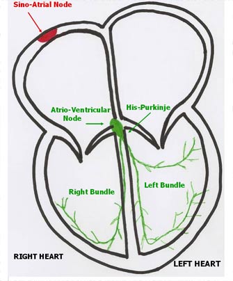
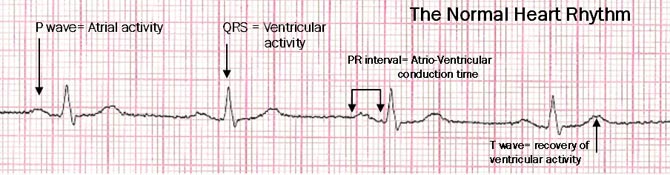
The heart pumps blood into a network of conduits called “arteries” to supply oxygen and nutrients to the body. The heart is a muscle that beats automatically, driven by an excitation-contraction sequence of muscle cells. Who is generating this sequence in the heart? A group of specialized cells drives the heart rhythm by generating spontaneous electrical activity, termed as the P wave on the electrocardiogram (EKG): these cells are named as the Sino-Atrial Node, and are located in the upper right chamber of the heart, the Right Atrium. Their pacemaker current spreads to the heart via a Conduction system of cells that have different electrical properties: the Atrio-Ventricular Node, the His-Purkinje, and the Branches (Figure 1).
A group of cells is named the “Atrio-Ventricular Node (AVN)”, as they are located across the upper chambers (the Atria) and the lower chambers (the Ventricles) of the Heart: here a conduction delay from the Atria to the Ventricles occurs, allowing contraction of the formers and proper filling of the latter. The His-Purkinje cells ensure fast conduction of the pacemaker current from the AVN to the ventricles, whose contraction elicits the strength to pump the blood into the arteries, supplying oxygen and nutrients to the whole body. The His-Purkinje system divides into two branches for each Ventricle, termed as the Right Bundle and the Left Bundle, respectively (figure 1). The conduction of the electric current to the ventricles causes a sharp deflection on the electrocardiogram, termed as the “QRS complex”, that is followed by recovery of ventricular excitability (the T wave). The recovery of Atrial excitability is “buried” within the QRS complex, and cannot be seen on the EKG because of its small amplitude.
The specialized cardiac cells taking part into the output and the conduction of the pacemaker current do not elicit muscular contraction.
The Sino-Atrial Node (SAN) cells have the greatest automatic pacemaker activity in the whole heart. They are connected the autonomic nervous system, that acts as a powerful modulator of cardiac activity, by either increasing or decreasing the heart rate according to the physiologic setting (physical or psychological activity, sleep, meals, emotional stress…) or to environmental factors ( temperature, altitude, depth ..).
The “normal” heart rate has a wide range, that changes according to age. Apart from trained sportsmen and physical workers, who may reach resting heart rates below 50 beats per minute (bpm), the average adult population has a resting heart rate between 50 and 90 bpm.
How can one detect the heart rhythm? The electrocardiographic recording (EKG) is the gold standard to detect and analyze the cardiac rhythm. However, one can easily measure the heart rate by the physical effect of the heart contraction: the arterial pulse. The arterial pulse is the mechanical propagation of the heart contraction, hence of the excitation-contraction sequence. The arterial pulse can be easily detected at the wrist (radial artery), at the neck (carotid artery), or at the groin (femoral artery).